January 07, 2021
Tag:
Sketch
Myelin oligodendrocyte glycoprotein is a myelin protein component that exists in the most superficial layer of the central nervous system myelin and is a subtype of immunoglobulin IgG1. Reasonable adjustment of complement-dependent cytotoxic response is an important component leading to MS demyelination.Anti-MOG antibodies play an important role in the pathological process of multiple sclerosis. The experimental immune cerebrospinal cord induced by MOG is an internationally recognized animal model of MS at this stage.

MOG polyclonal antibody
Physicochemical properties
MOG is present in the most superficial layer of the myelin membrane and oligodendrocytes, with a small number, accounting for about 0.01%-0.05% of the total production of myelin protein.Because MOG, compared with other myelin proteins, generally delays expression for 24-48 h during the developmental stage, it can be used as a marker to perfect oligodendrocytes.
Characteristic
1. Adhesion molecules
The OG immunoglobulin-like region and butyrophilin-like region were similar, and the chicken B-G antigen could not differentiate the effect of MOG.The surface localization of MOG, its understanding as an immunoglobulin superfamily, and the appearance of Lalpha/HNK-1 carbohydrate antigen epitopes all apply to the role of MOG as an adhesion molecule or cell receptor.Lalpha/HNK-1 epitopes found on MOG were evaluated as myelin-related glycoproteins and P0, both of which are myelin-specific proteins with adhesion components.If MOG has adhesion, it may have contact with other central nervous system components besides itself; it is inferred that if MOG has a significant role at the cell adhesion level, MOG may act as an adhesion "glue" between the adjacent myelin chemical fibers, resulting in the middle and late stages of the myelin production process.

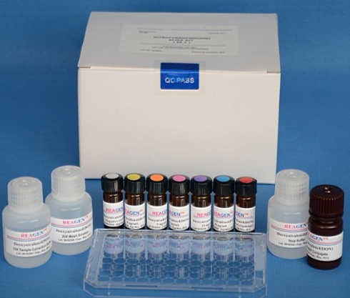
Mouse anti-myelin oligodendrocyte glycoprotein antibody (MOG-Ab) ELISA Kit
2. Activation of complement
The MOG gene is similar to the B-G antigen of chicken MHC at the site of the key institutional compatibility complex, and its structure is similar to the gene family, which can cause obvious immune response.It is inferred that MOG has immune functions in the central nervous system, and recent preliminary reports have reminded that MOG is expressed in a range of monocytes, which is also applicable to the immune function of MOG. Although MOG does not appear in non-feeding types of myelin, it can be expressed in several ways, one is that MOG does not directly act on the myelin process, more appropriately,It may mediate the contact between K myelin and another component of the body such as the immune system; another expression is that MOG is relatively specific to the immune function of CNS myelin, but not to the peripheral nervous system myelin, which can activate the classical complement mode in the body.Defensive DNA vaccine number MOG (MOG-DNA) can avoid the production of immune encephalomyelitis in the mouse experiment itself, which is completed according to the effect of complement C3d.Thus, CNS myelin must include complement components that fuse Ctq, leading to complement activation.
Mechanism of action
1. MOG and EAE
EAE is a comparatively ideal animal model for studying MS at home and abroad.At present, there are many reports about the establishment of EAE model with myelin as antigen at home and abroad. At first, the most used allergens are brain or spinal cord homogenate, MBP, PLP, MAG or its peptide brilliant fragments, etc.The sensitive species used are mostly rodents, such as Lewis rats, SJL/J mice, PL/J mice, SWXJ mice, etc. Others are monkeys, guinea pigs, rabbits, etc.Although MOG only accounts for 0.01% to 0.05% of the total myelin protein, it is highly antigenic.In the study of EAE, it was found that MOG is the only central nervous system myelin protein component that can cause both demyelinating antibody response and T cell response.Rats or mice immunized with MOG or its peptide fragments truly reproduced the present clinical history of multiple types of MS, including relapse-reduction (RR), primary progression (PP), secondary progression (SP), Devic's disease, and Marburg's disease.Biology has also confirmed that MOG is expressed on the surface of oligodendrocytes, inhibiting the regeneration of damaged axons according to fusion with the receptor NgR.MOG also exists on neurons, mainly long projection neurons, such as Purkinje cells of the thalamus, Fulmilla pyramidal cells, motor neurons of the spinal cord and anterior horn cells of the spinal cord.MOG is also involved in the regulation of cell proliferation, and overexpression of oligodendrocyte myelin glycoprotein in 3T3 fibroblasts has growth and development inhibitory effects.It can be inferred that MOG plays an important role in the pathogenesis of MS.

Myelin oligodendrocyte glycoprotein kit 1. Diagnosis of MOG and MS
2. MOG and MS
Immunologically CD4+ T cells with HLA-DR-restricted confounding advantages of antigenic epitopes are located in the transmembrane and intracellular parts of MOG, including 146-154 amino acids. Surprisingly, all normal control groups have a broader response to MOG peptides than MS groups.MOG transmembrane and intracellular fractions are more antigenic than extracellular fractions.In EAE models, antibodies to MOG can cause demyelination both in vivo and in vitro, whereas additional administration of anti-MOG antibodies can lead to progressive EAE models.Anti-MOG-Ig antibodies are prevalent in CNS inflammation.It is transient in OIND but persists in MS.Experiments confirmed that serum anti-MOG-Ig responses were established early in MS, and the frequency and titer were comparable in the middle and late stages of MS with those in the early stage.
Application of myelin oligodendrocyte glycoprotein
The positive rate of anti-MOG antibody in MS patients has a considerable degree of specificity, and it has been reported abroad that the positive rate is as high as 72%.It has been confirmed that gold immunolabeling of CNS tissues of MS patients has anti-MOG antibodies in their active demyelinating lesions, and MOG exists in the outermost layer of the myelin sheath, which is in direct contact with substances in the extracellular environment and is most vulnerable to invasion. Therefore, it is speculated that anti-MOG antibodies play an important role in the pathogenesis of MS.2. Prognostic anti-auto-MOG antibodies of MOG and MS may be biological diagnostic markers of MS.The predictive effect of antibodies was significant (P=0.004), but not according to McDonald's criteria.In addition, the data also revealed that antimyelin antibody-positive patients would relapse within a shorter time interval than negative patients.Although anti-MOG antibodies in individuals are associated with the risk of MS, this association is at least partially affected by MOG and EBNA cross-reactivity.
Write to the end:
Myelin oligodendrocyte glycoprotein peptides are used as organic and pharmaceutical intermediates. Go Top peptide Biotech Co., Ltd is a manufacturer specializing in peptide synthesis and peptide customization. It also supplies raw materials of myelin oligodendrocyte glycoprotein peptide. Companies that need to purchase APIs are welcome to purchase them.
For any requests of Peptide for research purpose, welcome to contact us. www.gtpeptide.com , sales1@gotopbio.com.


Contact Us
Tel: (+86) 400 610 1188
WhatsApp/Telegram/Wechat: +86 13621645194
Follow Us:


 Pharma Sources Insight January 2025


